Magnetic Resonance Imaging Features of a Case Series of Intraductal Papillary Neoplasms of the Bile Duct
Abstract
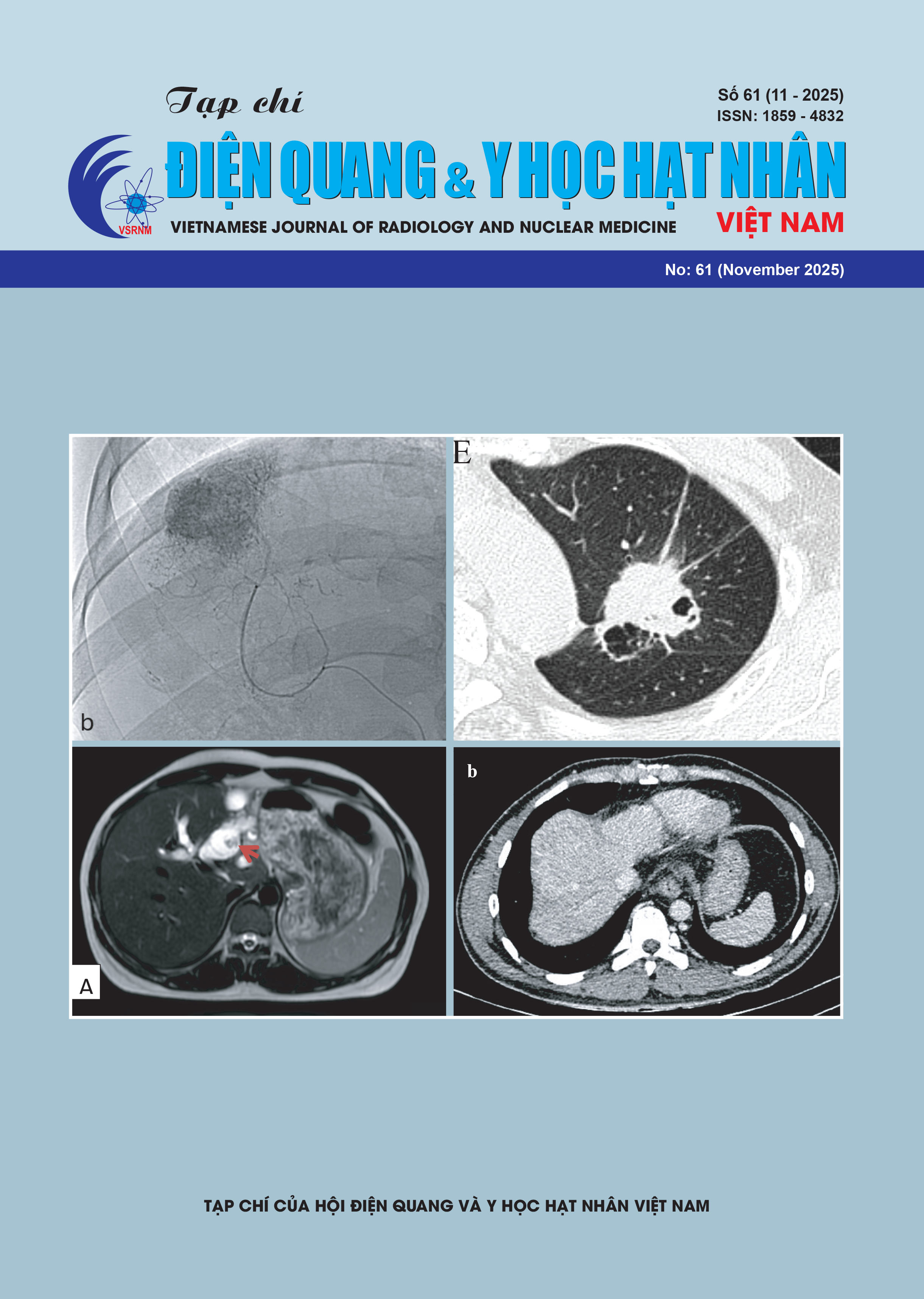
Background: Intraductal papillary neoplasm of the bile duct (IPNB) consists of papillary or villous proliferations of neoplastic epithelium within the bile duct lumen, supported by a thin fibrovascular stalk.
Case Presentation: We report five cases of IPNB that underwent magnetic resonance imaging (MRI) at Bach Mai Hospital. Clinical data, liver enzyme test results, detailed hepatobiliary MRI findings, and histopathological outcomes (from either biopsy or surgery) are presented for each case.
Conclusion: This case series further supports the notion that IPNB is a disease that can be diagnosed by MRI, featuring characteristic imaging patterns and morphological classifications that aid in deeper diagnostic orientation. The integration of imaging-based assessment and interventional planning plays a crucial role in the diagnosis, treatment, and prognosis of patients with IPNB.