RÒ ĐỘNG TĨNH MẠCH MÀNG CỨNG NỀN SỌ TẦNG TRƯỚC CÓ CẤP MÁU TỪ ĐỘNG MẠCH MẮT: BÁO CÁO 4 TRƯỜNG HỢP ĐƯỢC CAN THIỆP NỘI MẠCH
Tóm tắt
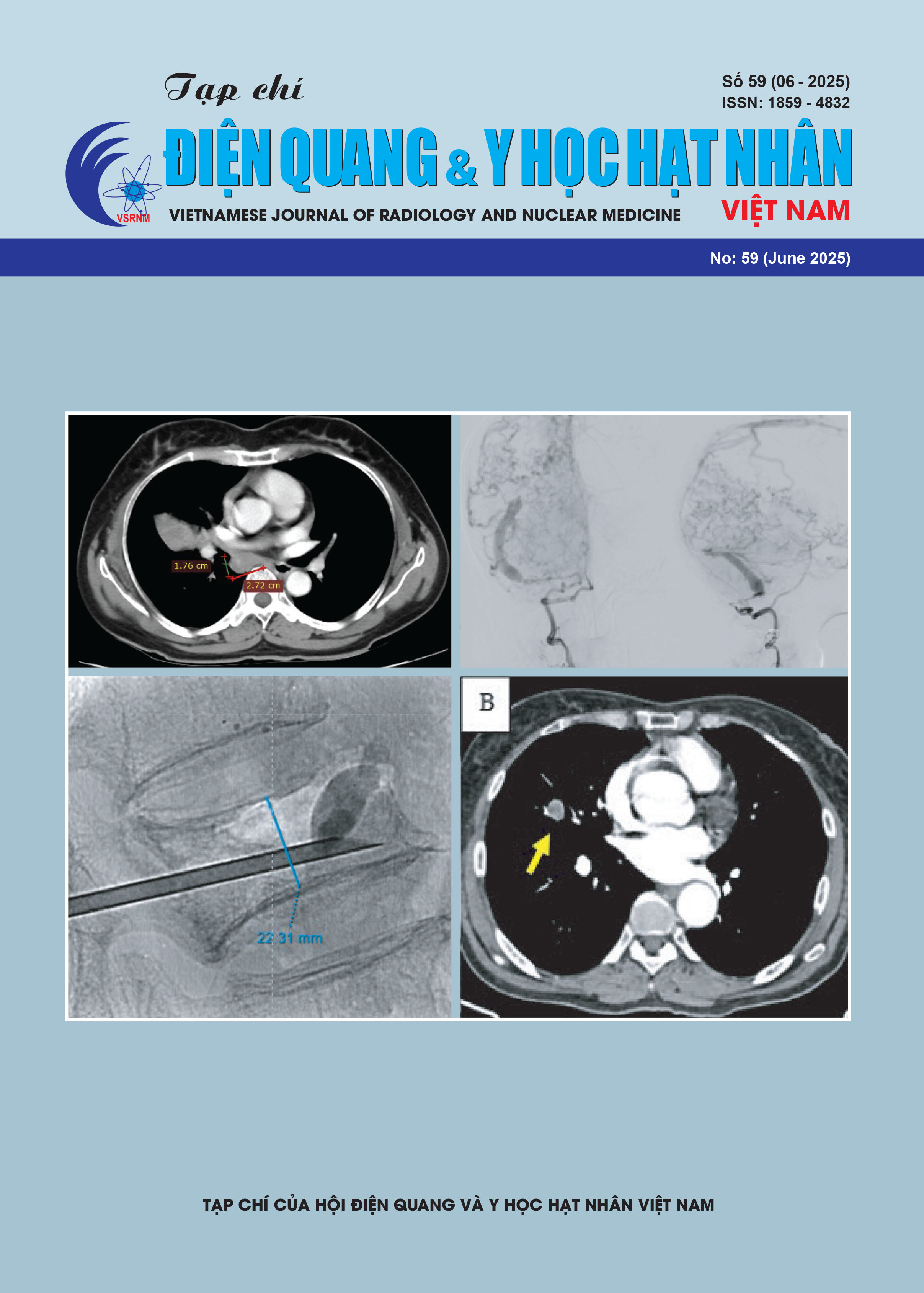
Mở đầu: Rò động tĩnh mạch màng cứng (RĐTMMC) vùng nền sọ tầng trước hiếm gặp với 5% so với RĐTMMC nói chung nhưng cơ xuất huyết cao hơn vị trí khác. Vị trí rò này thường có nguồn cấp máu từ nhánh sàng của động mạch mắt và do đó cách tiếp cận can thiệp nội mạch (CTNM) phải đảm bảo vừa tắc được vị trí rò vừa bảo tồn động mạch trung tâm võng mạc. Mục đích nghiên cứu là đánh giá hiệu quả và độ an toàn của can thiệp nội mạch điều trị RĐTMMC vùng nền sọ tầng trước, được cấp máu từ động mạch mắt.
Phương pháp nghiên cứu: Nghiên cứu hồi cứu 4 ca bệnh nhân rò vùng nền sọ tầng trước, được can thiệp tại Bệnh viện Chợ Rẫy từ tháng 1/2021 đến tháng 01/2024. Các đặc điểm lâm sàng và hình ảnh được đánh giá trước can thiệp, lúc xuất viện và tái khám sau 3 tháng.
Kết quả: Các ca bệnh có đặc điểm chung là vào viện vì giảm thị lực, 1 ca xuất huyết não và 1 ca lồi mắt, CT/MRI khẳng định nguồn cấp máu là từ nhánh sàng của động nhánh mắt với giãn lớn tĩnh mạch dẫn lưu. CTNM tiếp cận đường động mạch mắt trong 4 ca, trong đó 1 ca tiếp cận động mạch mắt hai bên, 1 ca tiếp cận nhánh màng não của động mạch cảnh ngoài để tắc bổ sung. Thành công kỹ thuật với tắc hoàn toàn vị trí rò và bảo tồn động mạch trung tâm võng mạc trong tất cả các ca. Vi ống thông được sử dụng là ống thông có đầu tách rời trong 3 ca, chất thuyên tắc Onyx 18 được sử dụng trong tất cả các ca. Biến chứng thủ thuật liên quan 1 trường hợp là xâm nhập onyx vào xoang dọc trên, tuy nhiên mức độ ít và không gây ảnh hưởng đến dòng chảy xoang tĩnh mạch dọc trên. Theo dõi sau can thiệp 90 ngày, không ghi nhận tái phát, cải thiện thị lực đáng kể với 3 trường hợp.
Kết luận: Can thiệp nội mạch tiếp cận đường động mạch mắt trong bệnh lý RĐTMMC vùng nền sọ tầng trước có khả năng cao tắc vị trí rò và bảo tồn động mạch trung tâm võng mạc.